Microscopy
Looking in on a huge tiny world!
I've always had a microscope, but its always been a cheap, toy type. Some have had metal bodies, but all have had horrible plastic optics, clunky focusing mechanisms, and wobbly stages to mount your slides on. My daughter Jen has recently become very interested in microbiology, so we decided to get a better microscope.
After doing some basic research, here is what I have identified as important features/aspects of a better microscope:
- glass optics (both eyepiece and objective)
- DIN objective lenses
- mechanical stage (focus shifts stage instead of objective lenses)
- coarse and fine focusing
- LED lighting
- metal body construction
 I am sure there are
more things someone would want in a microscope, but for me buying a unit for occasional use, even my
list turned out
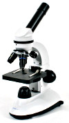
to be too much. In the end, I bought a My First Lab MFL-06 microscope from Mastermind Toys in
Barrie. At $89 (CAD),
this scope has glass optics, a mechanical stage, removable objectives (not sure if they are DIN or
not), coarse only
focusing and LED lighting. Fine focus would be nice, but we have had no problem finding a sharp
focus so far, and the
plastic body is quite high quality.
I am sure there are
more things someone would want in a microscope, but for me buying a unit for occasional use, even my
list turned out
to be too much. In the end, I bought a My First Lab MFL-06 microscope from Mastermind Toys in
Barrie. At $89 (CAD),
this scope has glass optics, a mechanical stage, removable objectives (not sure if they are DIN or
not), coarse only
focusing and LED lighting. Fine focus would be nice, but we have had no problem finding a sharp
focus so far, and the
plastic body is quite high quality.
So far, this little scope has vastly exceeded my expectations, providing crisp and clear images of everything we have looked at so far. It has a 10X eyepiece, plus 4X, 10X and 40X objectives providing powers of 40, 100 and 400. The 40X objective is of limited use, as it frequently crashes into the slide cover (there is no safety stop on the stage, and with only coarse focusing it is difficult to avoid!). The 10X objective is by far the most used, but it still provides amazing views of the microscopic world!
 We have added
a Celestron digital microscope imager
to our set up. It is a CCD camera with 2.0 megapixel resolution and an adapter to allow it to fit
just about any
microscope. It works perfectly with our MFL-06 scope. We have already captured some quality images
of microbial life.
We look forward to refining our skills with it and capturing some detailed images of the tiny
universe under our feet!
We have added
a Celestron digital microscope imager
to our set up. It is a CCD camera with 2.0 megapixel resolution and an adapter to allow it to fit
just about any
microscope. It works perfectly with our MFL-06 scope. We have already captured some quality images
of microbial life.
We look forward to refining our skills with it and capturing some detailed images of the tiny
universe under our feet!
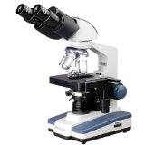
My next scope, I think, will be the AmScope B120B stereo compound microscope. It has all the features listed above, plus the advantage of stereo view. At $240 (CAD), it is a lot more money, but it is an entry level professional unit that will allow me to go to the next level in microscopy. The magnifications supported are 40X-2000X so I will be able to examine bacteria and other very small microbes. This scope is also very expandable (modular) so it will likely be the "last scope" I need to buy!
Microscopic Organisms
Here are some organisms that I have viewed through my MFL-06 microscope. Note: The identification of these organisms is just an educated guess on my part. If I have mis-identified anything here, please let me know at [email protected] and I will update this page. Thank you!
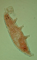 Milnesium tardigradum:
commonly called tardigrades or water bears, these tiny eight legged creatures range in size from
0.2mm to 0.7mm. They
fumble about through moss and other plant debris in search of food then they suck the fluids out of
plants and other animals.
Our examples appeared yellowish-brown when viewed in a water sample that was treated with blue dye.
They appeared very similar
in size to the nearby rotifers (see below), but they did not seem to bother with them. They really
do look like little
bears! [LINK]
Milnesium tardigradum:
commonly called tardigrades or water bears, these tiny eight legged creatures range in size from
0.2mm to 0.7mm. They
fumble about through moss and other plant debris in search of food then they suck the fluids out of
plants and other animals.
Our examples appeared yellowish-brown when viewed in a water sample that was treated with blue dye.
They appeared very similar
in size to the nearby rotifers (see below), but they did not seem to bother with them. They really
do look like little
bears! [LINK]
My notes:
- brown in colour
- same in size as the rotifers
- hooks visible on bottom of legs (feet)
- mouth pops out to eat
- crawls over/through plant debris
- does not seem to fight with rotifers
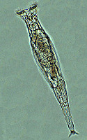 Rotaria rotatoria: commonly called
rotifers, these multi-cellular organisms are fascinating to watch. They have twin coronas with cilia
that move so fast, it
seems as though they are spinning blades or wheels. With these apparatus, the rotifers create a
vortex in the water they inhabit
so they can direct bacteria to their mouth which is located between the coronas. They are very
active, and in my latest water
sample (from Six Mile Lake Provincial Park in Ontario, Canada) they appear to be very common. Under
blue or red dye, they appear
brownish-yellow in colour, with a semi-clear outer skin, and mostly clear tail and foot (with
toes/claws).
[LINK]
Rotaria rotatoria: commonly called
rotifers, these multi-cellular organisms are fascinating to watch. They have twin coronas with cilia
that move so fast, it
seems as though they are spinning blades or wheels. With these apparatus, the rotifers create a
vortex in the water they inhabit
so they can direct bacteria to their mouth which is located between the coronas. They are very
active, and in my latest water
sample (from Six Mile Lake Provincial Park in Ontario, Canada) they appear to be very common. Under
blue or red dye, they appear
brownish-yellow in colour, with a semi-clear outer skin, and mostly clear tail and foot (with
toes/claws).
[LINK]
My notes:

- yellow-brown in colour
- clear tail
- rotors create vortex to suck in food
- seems to like eating paramecium?
- also eats plant debris?
- moves by planting it's tail, stretching out, grasping with head, then squishing up (like a worm)
- rotors are fantastic & deadly! (not really, but they look SO cool! actually waving cilia)
- do not seem to fight each other or neighbouring tardigrades
- can swim in open water fairly quickly
- heart beats 3 times, takes a break, then 3 times again
- size: 1/5th width of field of view at 100x
- immature rotifers are clear with no rotors and slimmer than adults?
 Chlamydomonas reinhardtii: also
known as green algae, these are single-celled organisms that are about 10 micrometers in diameter.
It has two small
flagella that it uses to move around. It does not move very quickly, but I can see them move in my
microscope. They are
bright green with a visible clear(ish) membrane. They have a squared-off oval shape, that sometimes
turns round and goes
back to oval. Not sure if that's an actual shape change, or just a perspective change as it shows a
different aspect?
[LINK]
Chlamydomonas reinhardtii: also
known as green algae, these are single-celled organisms that are about 10 micrometers in diameter.
It has two small
flagella that it uses to move around. It does not move very quickly, but I can see them move in my
microscope. They are
bright green with a visible clear(ish) membrane. They have a squared-off oval shape, that sometimes
turns round and goes
back to oval. Not sure if that's an actual shape change, or just a perspective change as it shows a
different aspect?
[LINK]
My notes:
- bright green
- clear membrane
- circles inside
- move, but very, very slowly
- squared-oval in shape, sometimes turning round?
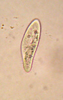 Paramecium aurelia: is a
common protist that has shape similar to the sole of a shoe. It was one of the first microscopic
organisms studied by our
species. This was also one of the first microscopic organisms that I studied as a student. It was
fascinating to see an animal
that was so different from us, yet functioned in some similar ways. Eating and digestion are
certainly obvious functions we
share, as well as reproduction. To see them scoot around under the microscope is one of the things
that hooked me on my love
of science! [LINK]
Paramecium aurelia: is a
common protist that has shape similar to the sole of a shoe. It was one of the first microscopic
organisms studied by our
species. This was also one of the first microscopic organisms that I studied as a student. It was
fascinating to see an animal
that was so different from us, yet functioned in some similar ways. Eating and digestion are
certainly obvious functions we
share, as well as reproduction. To see them scoot around under the microscope is one of the things
that hooked me on my love
of science! [LINK]
My notes:

- clear (no obvious colour unless a dye is used)
- very fast swimmers (compared to rotifers)
- seem to spiral in order to swim?
- change direction randomly
- seem tasty to rotifers?
- never seem to stay still!
- shaped like the sole of a shoe
 Spirogyra (?): This is certainly some kind of green algae in the form of a long tube. You
can see a split or division
in the tube near the top of it. I suspect it is a species of Spirogyra, but I cannot confirm a
spiral form to the chloroplasts
inside the tube. I realize this snapshot is a bit fuzzy - we are still working out the operation of
our digital microscope
eyepiece! Click on the image to the right to get a better view. [LINK]
Spirogyra (?): This is certainly some kind of green algae in the form of a long tube. You
can see a split or division
in the tube near the top of it. I suspect it is a species of Spirogyra, but I cannot confirm a
spiral form to the chloroplasts
inside the tube. I realize this snapshot is a bit fuzzy - we are still working out the operation of
our digital microscope
eyepiece! Click on the image to the right to get a better view. [LINK]
My notes:
- long thin tube/membrane
- mottled green contents (chloroplasts?)
- does not move (that I could see)
 Closterium navicula:
This algae moves at a moderate pace as it glided across my slide in a stately fashion. Usually
Closterium are crescent shaped,
but this species is boat shaped, with a conspicuous vacuole at each end. In my image, it is hard to
separate the two chloroplasts,
which are separated by the nucleus (but it is there!). Some other spheres are visible, which are
likely pyrenoids.
[LINK]
Closterium navicula:
This algae moves at a moderate pace as it glided across my slide in a stately fashion. Usually
Closterium are crescent shaped,
but this species is boat shaped, with a conspicuous vacuole at each end. In my image, it is hard to
separate the two chloroplasts,
which are separated by the nucleus (but it is there!). Some other spheres are visible, which are
likely pyrenoids.
[LINK]
My notes:
- about the same size as a rotifer
- moves in a straight line (no zig-zagging)
- vacuoles very visible at ends
- hard to discern delineation between chloroplasts and nucleus
- very green in colour (with some yellowing towards the middle)
- visible outer membrane appears darker (almost brown)
 Navicula lanceolata:
This fresh water diatom (phytoplankton) was gliding past a pandorina of some sort. It has a
distinctly brown tint to its green colour.
My example has some visible activity inside the membrane with green and brown spheres present.
[LINK]
Navicula lanceolata:
This fresh water diatom (phytoplankton) was gliding past a pandorina of some sort. It has a
distinctly brown tint to its green colour.
My example has some visible activity inside the membrane with green and brown spheres present.
[LINK]
My notes:
- top (value) view of diatom (side/girdle view would be rectangular)
- brown / green in colour
- moves at slow pace
- travels in a straight line
- does back up, then go forward again